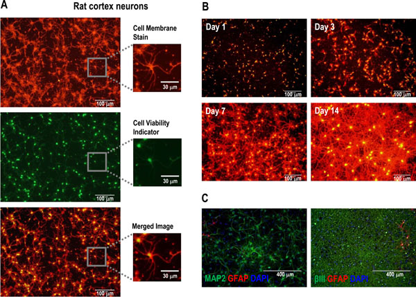
Fig. (2) Multiplex fluorescent dye-based staining of neurite outgrowth and neuronal cell health. (A) A 7-day culture of rat cortex neurons
was stained using an orange-red fluorescent Cell Membrane Stain, which serves as a reporter of neurite outgrowth, along with a greenfluorescent
Cell Viability Indicator that requires intracellular esterase activity. (B) Cryopreserved rat cortex neurons were plated and cultured
under neurite outgrowth-promoting conditions, then co-stained with the Cell Membrane Stain (orange) and the Cell Viability Indicator
(green) at different time-points. (C) Immunocytochemistry analysis of the rat cortex neuron cultures for neuronal markers MAP2 and beta-III
tubulin and glial marker GFAP.