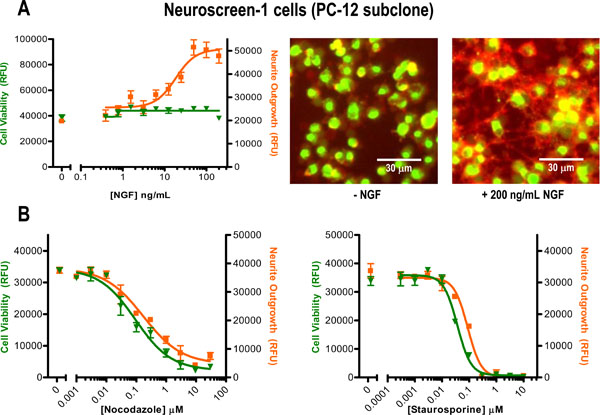
Fig. (3) Plate reader quantification of relative neurite outgrowth and cell viability using Neuroscreen-1 (PC-12 subclone) cells. (A) Cells
were plated in 96-well format and treated with a serial dilution of nerve growth factor (NGF) for 4 days. Prior to staining them, the cells were
fixed with 4%formaldehyde to improve their adherence to the assay plate. Relative cell viability (▼) and neurite outgrowth (■) were measured
using a bottom-read fluorescence microplate reader to detect the green and orange-red fluorescence intensity from each well. Data plotted
are the mean ± SEM (n = 4 replicate wells) of one representative experiment of three independent experiments that were performed. For
visual reference, representative merge images are also shown. (B) In a separate experiment, cells were plated in the presence of 100 ng/mL
NGF and then treated with titrations of known cytotoxic compounds Nocodazole and Staurosporine for 3 days (data are mean ± SEM, triplicate
wells).