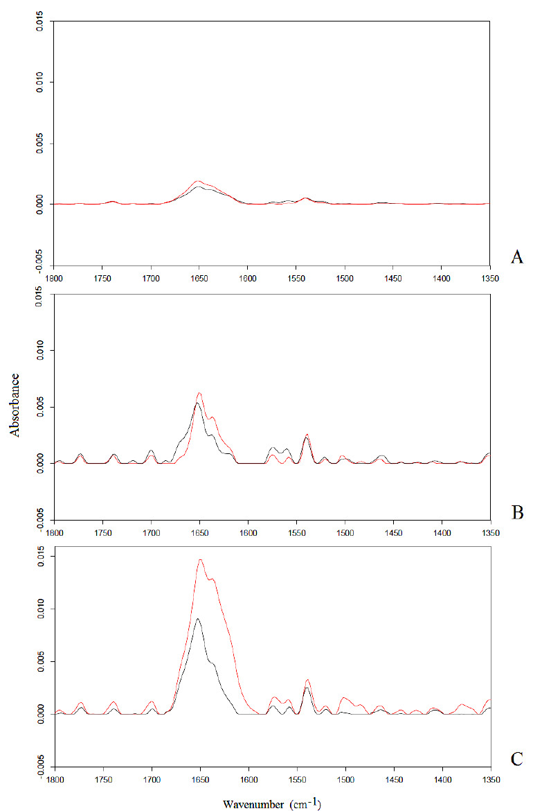
Fig. (1) Amide I band in neuronal-like cells after 6 h exposure to a static magnetic field (A), 50 Hz (B) and 900 MHz (C) electromagnetic fields. Spectra of exposed and unexposed samples are represented in the colors red and black, respectively. The increase in the intensity of the Amide I band with an increase of the frequency of the applied electromagnetic field after exposure can be observed, which can be explained assuming that α-helices in cells membrane channels had aligned towards the direction of the applied field.