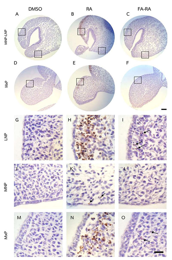
Fig. (3)
TUNEL assay of embryos treated with DMSO, 13-cis-RA (RA) and a combined folic acid and 13-cis-RA (FA-RA). The 13-cis-RA treated embryos showed numerous TUNEL-positive cells (brown staining) in the mesenchyme adjacent to the epithelium of dorsolateral part of LNP and MxP, (B, E, H, N) compared to those of the DMSO-treated embryos. A lower amount of positively stained cells found in 13-cis-RA treated embryos noted in the MNP (K, arrows). A dramatic reduction of TUNEL-positive cells was particularly found in the LNP and MxP of FA-RA-treated embryos (4 C, F and I, O arrows) compared to those observed in the 13-cis-RA treated embryos. G-O showed a higher magnification of square area in A-F. Scale bar= 100 µm for A-F and 20 µm for G-O.