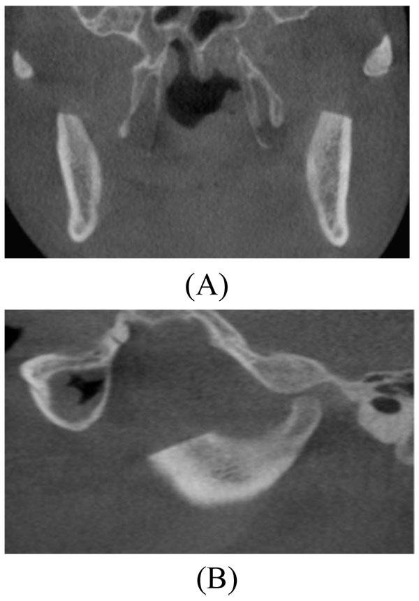
Fig. (8)
Postoperative Cone Beam Computed Tomography scan. (A) Coronal image showing no interference between the coronoid processes and the medial surface of the zygomatic bone and arch. (B) Sagittal image showing the resected coronoid process with no interference with the zygomatic bone.