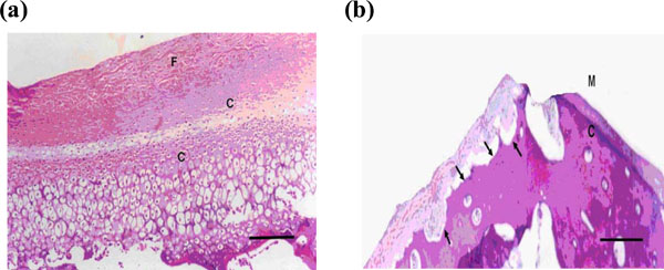
Fig. (3) (a) Microphotograph of the condyle after 2 weeks showing signs of active growth and increased thickness of the fibrous layer and hypercellularity of the chondrocytic layers, which was noticed only in the middle and posterior areas of the condylar surface (H & E X100). (b)Microphotograph of the condyle by the end of the 2nd week showing signs of bone resorption and evidence of repair with fibrous tissue, at the anterior articulating area of the condyle (H & E X 40).