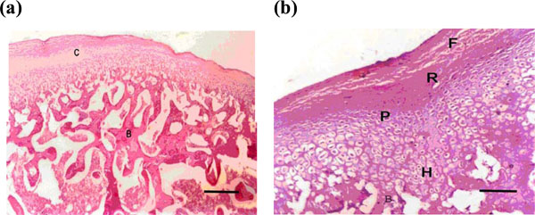
Fig. (4) (a) Microphotograph of the condyle after 3 weeks showing hypertrophy of the cartilaginous layers and reformation of many bone trabeculae (H & E X100) (Bar = 100 micrometer). (b) Higher magnification of photo no 4a (H & E X200) (bar = 200 micrometer).