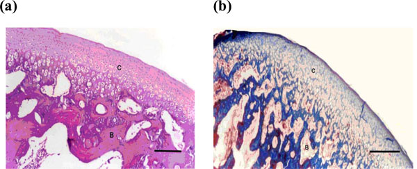
Fig. (5) (a-b). Microphotograph of the condyle after 4 weeks showing histologic features of the anterior part of the condyloid process that looks nearly normal after regeneration of all layers of the articulating cartilage (C) and the subcartilaginous bone (B) (a = H & E X200, b = Mason Tri-chrome X200) (bar = 200 micrometer).