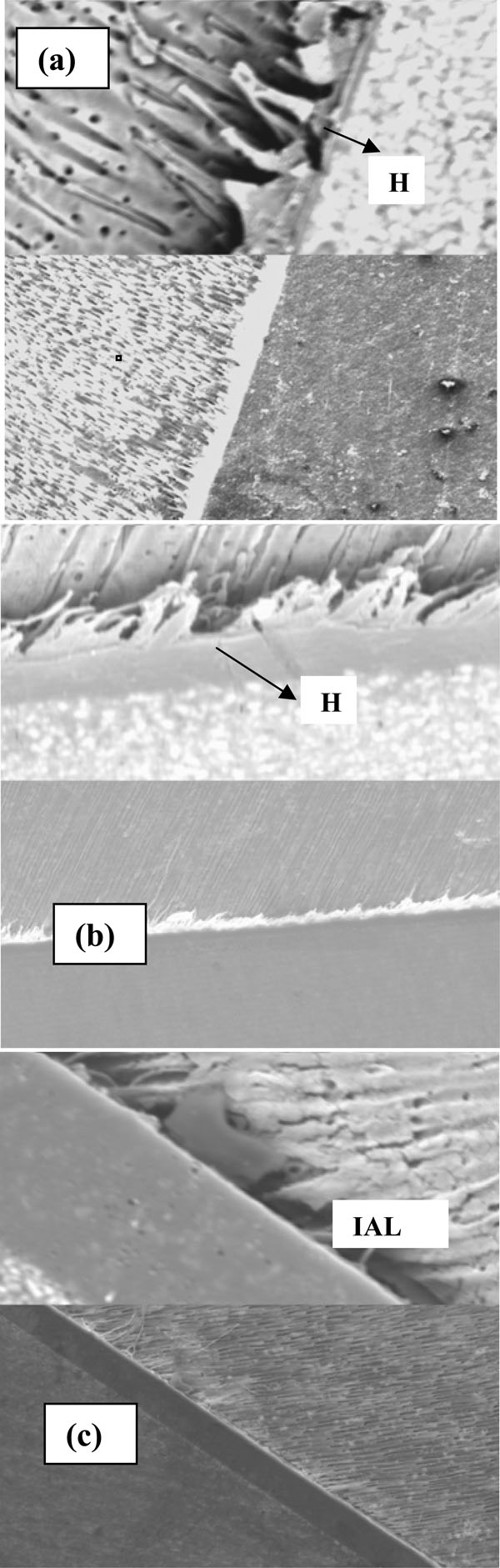
Fig. (3) Split micrographs (200X bottom/1000X top) of dentin-adhesive interfacial morphology (a) PNB (b) SEB and (c) OUP. The hybrid layer is marked ‘H’ in all images. Note the distinct hybrid layer morphology in PNB and SEB. In OUP, a thick interfacial adhesive layer (IAL) appears to have fully dissolved the smear layer including smear plugs into the tubules.