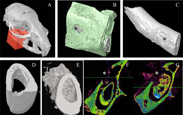
Fig. (3) Micro-scanner analyzes of incisor implanted with the 17IA4 cells. The red block indicates the zone analyzed (A) and the asterisk(*) corresponds to the perforation site in the bone (B), and in the tooth (C). Ten days after implantation in the pulp, no mineralization was observed in the control group (D,F) in contrast, the 17IA4 group formed a large mineralized structure within the pulp (E,G).