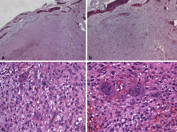
Fig. (6) Histological features of the tissue stained with haematoxylin-eosin. (a) Low magnification (E-E, 25X) figure showing granulation tissue and bone islets. (a) Higher magnification (E-E, 50X) of the previous slide. (c) Tissue section illustrating the detailed histomorphology of the lesion (E-E, 200X). (d) High resolution image (E-E, 400X) of multinucleated giant cells.