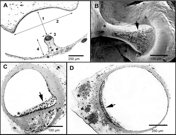
Fig. (3) Optical and scanning electron micrographs of the axolotl inner ear. In A, section of the posterior SC ampulla. The ampulla is delimited by the narrowing of the SC. Crista ampullaris is located in the midst of the ampulla. Figure shows the measurement of: 1- ampulla heigth (2a0), 2- ampulla length, 3- crista thickness (2δ), 4- crista height. Mielinated axons innervating the crista hair cells are identifiable (*). In B scanning electron microscopy and in C, optical microscopy of a transversal section of the lateral semicircular crista ampullaris. This is a non symmetric structure with a large widening in the external wall of the ampulla (arrowhead) upon which hair cells accumulate, and leaving what seems to be a cleft in the internal side of the ampulla (*). Pictures in B and C are from different animals. In D, cross section of the utricle. The macula (arrowhead) is located ventrally and occupies about a third of its perimeter. Mielinated axons innervating the macula hair cells are located beneath the neuroepithelia forming three rami (*).