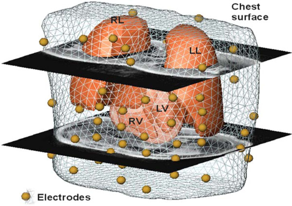
Fig. (1) Volume conductor model generated from 3D cine MRI data with two axial MR scans with overlaid electrode positions (segmented liquid capsules) from a left lateral oblique view; also shown: the triangular model of the chest surface, the surface models of the both lungs (LL, RL), and the ventricular model (RV, LV).