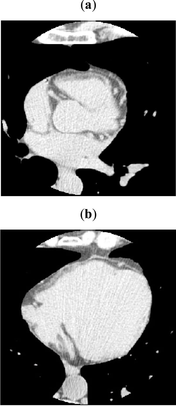
Fig. (1) Typical slices acquired in the calcium scoring case. In the slice (a) some portion of atria and great vessels are visible, while slice (b) shows a separation problem between the heart and the chest wall. Hounsfield values between and are represented.