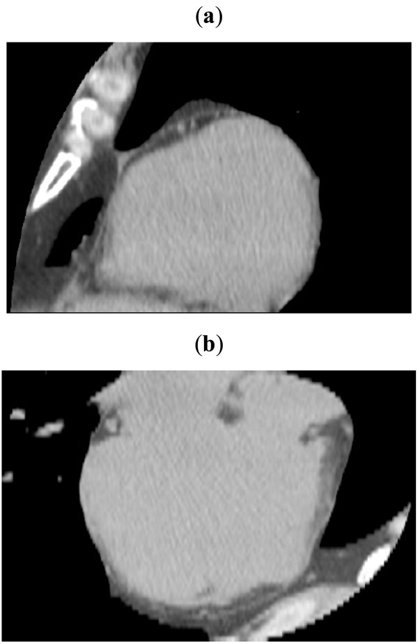
Fig. (2)
Typical views of the dataset obtained after resampling in planes parallel to the atrioventricular sulcus: (
a
) saggital view and (
b
) axial view. Notice that in (
a
) the atrioventricular sulcus is easily discernible.