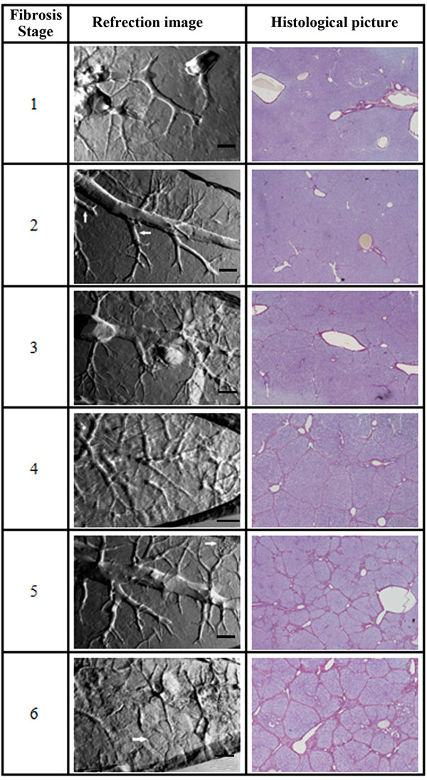
Fig. (3)
The refraction images of hepatic fibrosis samples and the corresponding histological images from stage one to six. The bars in the refraction images are 500 µm.