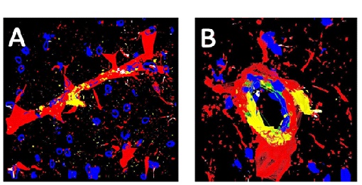
Fig. (5)
To have a structural view of eNOS (red) with vessels, we made rotatable, 3D models Fig. (5), showing eNOS around a venule (panel A) and an arteriole (panel B) in a section of olfactory bulb, demonstrating specificity of this NOS isoform to endothelial cells. Smaller vessels can also be seen coursing throughout the tissue. Connective tissue staining has been removed for clarity (Magnification x 1200).