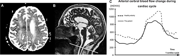
Fig. (1) A: The T2* Magnetic Resonance Imaging shows three areas of semi recent ischemia in the white matter, including: frontal right posterior, right posterior transitional zone and beside the left central sulcus.
B: A sagittal T2 sequence was used as localizers to select the anatomical levels for the Phase Contrast Magnetic Resonance Imaging used for the flow quantification over the cardiac cycle (CC). Acquisition plane was selected to be perpendicular to the vertebral and internal carotid arteries.
C: The graph shows the curves of the mean cerebral arterial blood flows, for the patient and a healthy elderly subject. This quantitative acquisition shows a decrease of pulsatility of the arterial cerebral blood flow of the patient over the cardiac cycle.