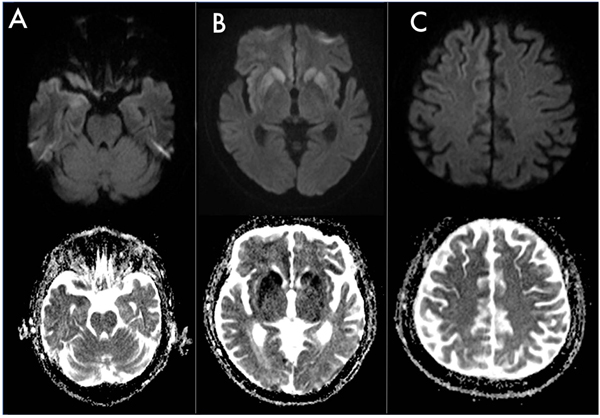
Fig. (1) MRI features. DWI (b1000) and ADC map: brainstem (A); basal ganglia (B); frontal paramedian cortex (C). No signal abnormalities are detectable in the brainstem. Note the marked DWI hyperintensity-ADC hypointensity involving putamen, caudate nuclei and right insular cortex (B), more subtle involving frontal paramedian cortex (C). Images are shown in radiological convention.