Fig. (2)
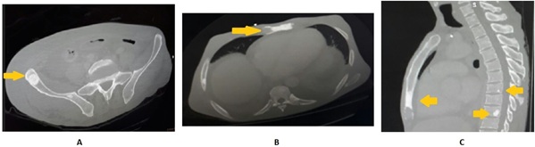
CT images showing sclerotic bone lesions in the right ilium
(a)
, lower third of the sternum body
(b)
, and thoracic vertebrae
(c)
. Lesions exhibited the typical pouch and soup-bubble-like appearance with a clear sclerotic rim (arrows).