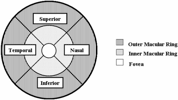
Fig. (1)
Optical coherence tomography macular map display, right eye. The macular map is divided into outer and inner rings, as well as four quadrants: superior, nasal, inferior, and temporal.