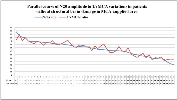
Fig. (11) Couples of N20 and 1/vMCA ratio values detected in patients of group 1 and group 2, who had no CT/MRI image of structural ischemic or haemorrhagic injury in MCA supplied area, displayed in descending order. Modifications of interhemispheric ratio of 1/vMCA correspond with analogue variations in interhemispheric ratio of N20 amplitude in a wide range of values, from 0.9 to 0.4-0.3, (p<0.01, r = 0.93, Cl for r = 0.89 to 0.96 unless structural ischemic or haemorrhagic damage develops.