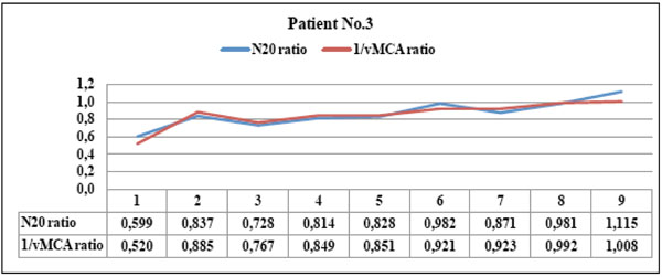
Fig. (5) Patient No. 3 showed parallel courses of N20 and 1/vMCA interhemispheric ratios with significant statistical correlation ( p < 0.01, r =0.91, C.I. 0.64 to 0.98). The patient showed TC scan image of parietal oedema corresponding to both ratio values <0.65 in the first examination (point 1); this promptly recovered to ratio values >0.65 after oedema disappeared (point 2). This patient never reached the vasospasm threshold, always maintained N20 amplitude > 1,2 µV on the compromised hemisphere and showed clinical evidence of lateralizing strength deficit with respect to the first examination only (point 1).