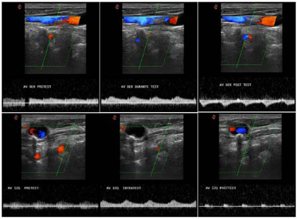
Fig. (1) Extracranial color Duplex study of vertebral arteries before (left side), during (middle position), and upon completion (right position) of the arm compression test. Right VA (upper images) reveals initial anterograde flow that became retrograde after cuff release. Left VA (lower images) also shows initial anterograde flow with loss of diastolic flow at the end of the test.