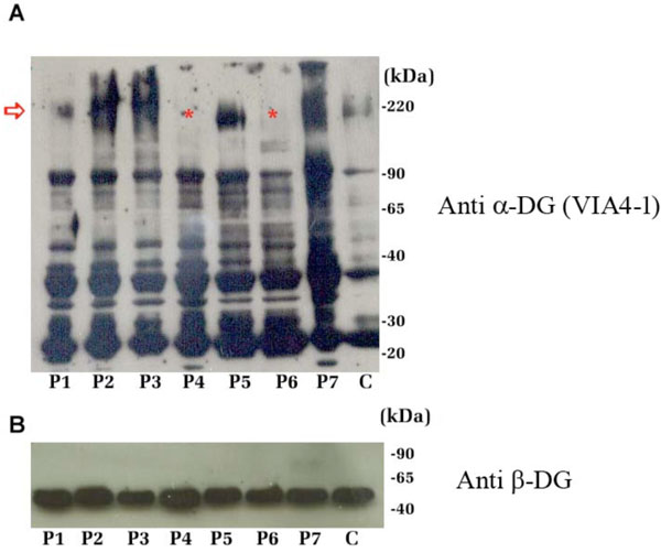
Fig. (3) Western blot analysis of the DG subunits in total protein extracts from patients P1-P7 and from control muscle tissue (C). A: Samples were resolved with a 7.5-15% acrylamide gradient SDSPAGE and α-DG was detected with the antibody VIA4-1 (lot 32685) raised against an undefined carbohydrate epitope (red arrow). In samples P4 and P6 the expected 156 KDa band was absent (red asterisk). The additional lower bands detected in all the samples might correspond to hypoglycosylated and/or proteolytic forms of α-DG; B: Western blot upon a 12% SDS-PAGE of the total protein extracts using anti 43DAG/8D5 for β-DG detection. All the samples showed the expected 43 kDa band.