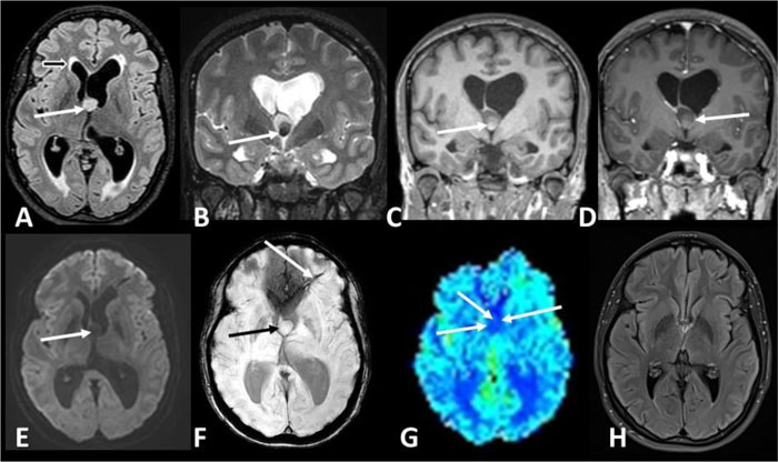
Fig. (1)
Axial FLAIR image (A) demonstrates a hyperintense lesion in the foramen of Monro (white arrow A), causing obstructive hydrocephalus. Hyperintensities in the periventricular white matter are attributed to periventricular interstitial edema (black arrow A). Coronal T2 image (B) reveals a hypointense area in the lower central part of the cyst (arrow B), which is hyperintense on T1 images (arrow C). Minimal peripheral enhancement (arrow D) is noticed on the coronal T1 image post gadolinium administration and it is attributed to the enhancement of the adjacent stretched septal veins. No diffusion restriction was demonstrated on DWI (arrow E). SWI showed neither microbleeds nor calcifications in the mass (black arrow F). A developmental venous anomaly in the left frontal lobe shown (white arrow F) was a clinically insignificant, incidental finding. Low rCBV was noticed on DSC-PWI cerebral blood volume map (arrow G). Follow up FLAIR axial image one year after the resection (H) demonstrates the normal appearance of the lateral ventricles, without transependymal edema.