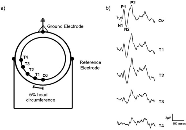
Fig. (1) (a) The electrode montage used to record motion-onset VEPs from the human scalp. Starting from Oz, each of the lateral electrodes (T1-T4) were placed at intervals equal to 5% of the observers’ head circumference. The ground electrode was placed on the forehead and linked ear electrodes acted as reference. (b) Sample motion-onset VEPs recorded from a single subject which were elicited by a moving high contrast L-M chromatic stimulus (10 deg/s) recorded from the five different electrode sites (Oz - T4).