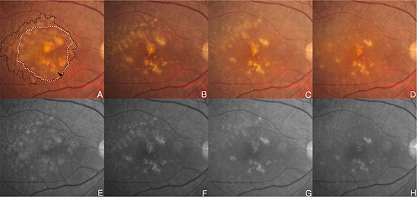
Fig. (2)
(A, E) Initial examination, fundus photography and red free fundus image in the right eye revealed DPED (white dotted line) with diffuse coalesced soft drusen (black dotted line) at macular and paramacular region. There are focal hyperpigmentary changes and retinal hemorrhage (black arrowhead) over confluent drusen.
(B, F) Six months after 5 intravitreal ranibizumab injections in the right eye, DPED at foveal area and drusens located superotemporal to macula are diminished over time. Retinal hemorrhage have also disappeared.
(C, G) One month after the initial absorption of drusen and DPED, drusen and central DPED reduction continued.
(D, H) Two months after initial absorption of drusen and DPED, previous central DPED reduction continued, and drusen at the superotemporal to macula area was decreased. But, CNV was recurred.