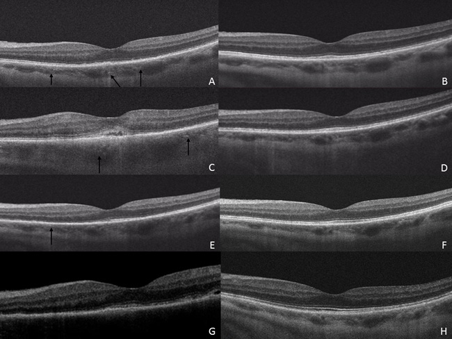
Fig. (1)
Optical Coherence Tomography (OCT) of each separate occurrence at presentation and at follow-up after resolution. (A) OCT of initial episode showing diffuse loss of the photoreceptor layer centrally (arrows) with choroidal thickening (B) OCT after vision recovery two weeks later showing recovery of the photoreceptor layer. (C) OCT on presentation after sudden vision loss eleven weeks after first episode. Note the diffuse loss of photoreceptor layer temporally (arrows) and serous detachment under the fovea. The second flare shows choroidal thickening, subretinal fluid, punctate hyperreflective dots (arrows) and outer photoreceptor disruption (C) with spontaneous improvement (D) one month later. The third flare shows choroidal thickening, punctate hyperreflective dots (arrows) and loss of the ellipsoid layer in the nasal macula (E) with spontaneous recovery (F). The fourth flare displays outer photoceptor loss (G) with resolution after systemic treatment (H).