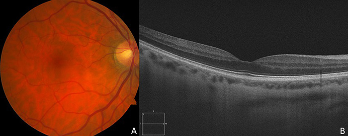
Fig. (2)
Color fundus photograph (
A
) and OCT (
B
) of the uninvolved right eye. Note the lack of inflammation or edema in the color photograph. Note the normal and intact photoreceptor layer on the OCT.