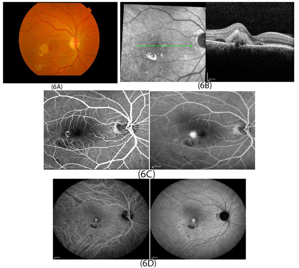
Fig. (6)
Multimodal imaging of type 3 CNV in patients with wet AMD. Fundus photography (Fig. 6A) of the right macula shows small intraretinal hemorrhage temporal to the foveal center with hard exudate inferior to the hemorrhage. OCT image (Fig. 6B) shows retinal thickening with intraretinal and subretinal hyperreflective materials and subretinal fluid. Fluorescein angiography (Fig. 6C) shows leakage of the right-angle vessels, which are temporal to foveal center and the blockage of the choroidal flush inferior to the leakage. ICG angiography (Fig. 6D) shows hypercyanescence and late leakage from abnormal vessels.