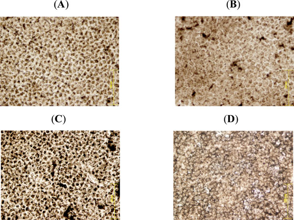
Fig. (4) Staining for lens epithelial ATPase activity. (A) Control lens epithelium shows a homogenous cell layer with brown spots of ATPase activity at the cell membrane. (B) Exposure to 90 cycles of microwave electromagnetic radiation caused some changes in the cells, there are spots of increased ATPase activity and the cells are larger than control cells. (C) 192 cycles of electromagnetic radia-tion caused dramatic increase in ATPase activity, with abnormal cell morphology and dead cells with no ATPase activity. (D) Con-ductive heat-treated lenses. Treatment of lenses at 39.5°C caused the epithelial cells to swell. ATPase activity could not be detected. Scattered between the swollen cells were small cells with high AT-Pase activity. Comparison between (C) and (D) demonstrates that the effects of micorwaves on the lens epithelium are remarkably different from the effects of heat.