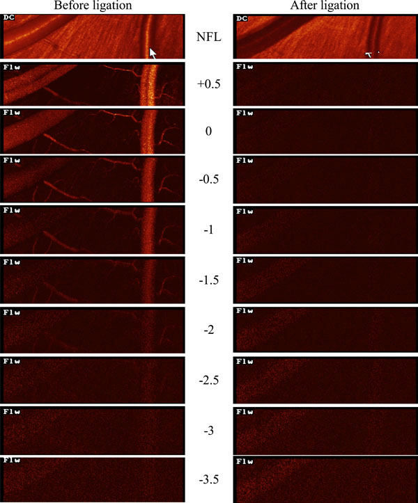
Fig. (1) HRF flow maps before and after ligation of retinal arteries. Ophthalmic Artery cannulated, perfusion rate = 500 µL /min. Before ligation of the retinal arteries the retinal vasculature is clearly evident in the HRF flow maps when the focal plane is in the superficial retina (upper left panels). At deeper focal planes the retinal vasculature becomes less evident but there is still no pattern associated with the choroidal vessels in the HRF flow map. After ligation of the retinal arteries (right panels), the retinal vessels are no longer evident in the HRF flow map, and again there is no evidence of a choroidal flow pattern in the deeper focal planes.