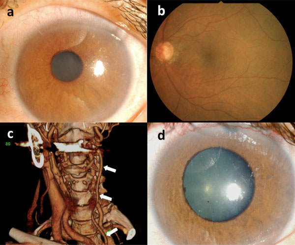
Fig. (1) (a) Slit-lamp examination showed corneal edema and rubeosis iridis in the left eye. (b) Fundus examination showed glaucomatous changes of the optic disc and almost normal retinal vessels. (c) Three-dimensional computed tomography angiography revealed absence of the left internal carotid artery and thinned common carotid artery (arrows). (d) A few days after the trabeculectomy, rubeosis iridis disappeared.