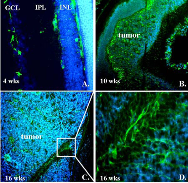
Fig. (1) VEGFR-2 immunofluorescence staining in LHβTag transgenic retinoblastoma. Representative eyes from LHβTag mice at 4 (A), 10 (B), and 16 (C,D)weeks are shown. VEGFR-2 immunoreactivity is green and DAPI counterstaining is blue. GCL, ganglion cell layer; IPL, inner plexiform layer; INL, inner nuclear; ONL, Outer nuclear layer. Magnification (A) 200 X, (B) 100 X, (C)200 X. (D) zoomed image from inset of C.