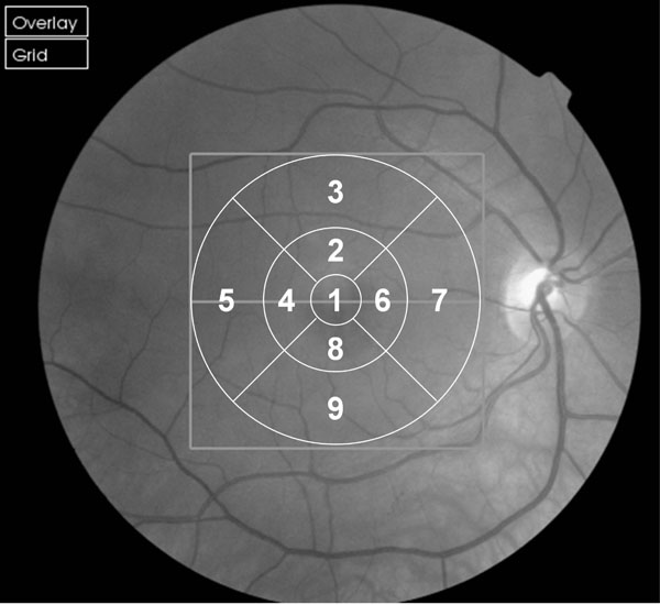
Fig. (1)
Fundus image of right eye with overlaid ETDRS 9 Region Map. Regions numbered for use in data analysis. For left eyes the region numbers were horizontally mirrored to maintain naso-temporal classification.