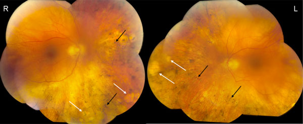
Fig. (1)
Fundus photographs. The color funduscopy of the right eye (R) and left eye (L) show blunted foveal light reflex, peripheral “punched out” lesions (white arrows) and bone spickling lesions (black arrows) bilaterally.