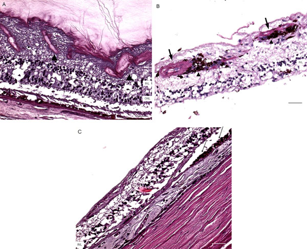
Fig. (4) Microscopic retinas. A, some small retinal vessels are sclerotic (arrowheads) in the right eye. B, some occluded vessels (arrows) are surrounded by pigment granules and a few migrated RPE cells (arrowheads) in the right eye. C, The left eye shows an absence of choriocapillaris, RPE, photoreceptors and the outer nuclear layer, while the remaining inner nuclear layer was adherent to an irregular Bruch’s membrane. Hypertrophic RPE (arrows) was present at the margin of these lesions. (GCL, ganglion cell layer; INL, inner nuclear layer; ONL, outer nuclear layer; scale bar indicates 50 µm).