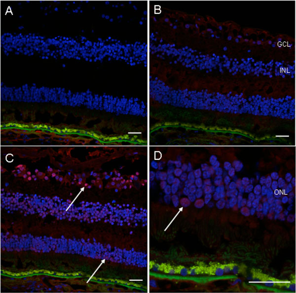
Fig. (6) Immunofluorescence detection of autoantibody in the patient’s serum on the normal retinal sections. A, no serum incubation (control); B, normal human serum incubation (1: 100, control); C, patient’s serum incubation (1: 100); D, higher magnification photo of C. There is positive fluorescence in the ganglion cell layer, inner nuclear layer and outer nuclear layer (white arrows) of the normal human retinal section incubated with the patient’s serum (Blue fluorescence = nuclear, DAPI staining; green fluorescence = autofluorescence; red fluorescence = positive staining). (GCL, ganglion cell layer; INL, inner nuclear layer; ONL, outer nuclear layer; scale bar indicates 50 µm).