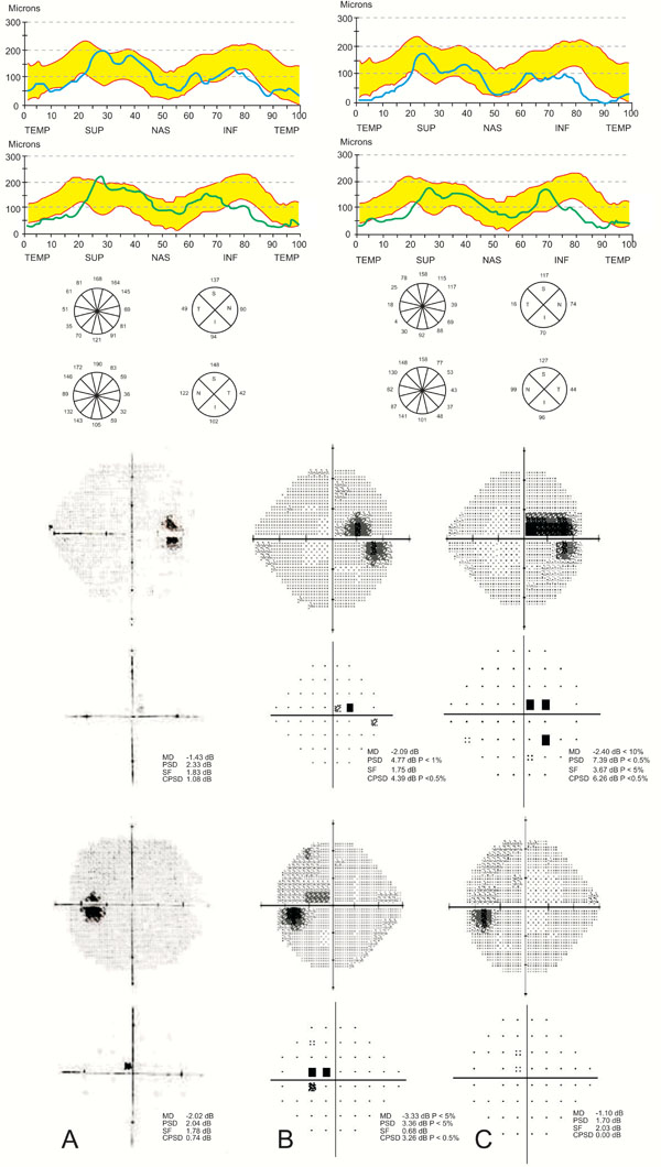
Fig. (2) (A) Top: The mean initial OCT RNFL examination of 5 scans is presented in clock-hours, quadrants, and graphically, in
comparison with the OCT age-matched normative database software provided by the manufacturer (bound in yellow). The RNFL is thinner
than the lower normal border in both the right (OD) and left eye (OS) superiorly, inferiorly and temporally. Bottom: Standard visual fields
are normal. (B) Visual fields in September 2007. Cecocentral scotoma is evident bilaterally, more in the left eye. (C). Top: In May 2008,
further thinning of the RNFL is detected in the inferior, superior and temporal quadrants of the right eye and in the superior quadrant of the
left eye. Bottom: The cecocentral scotoma widened in the right eye and became non-specific and less distinct in the left eye.