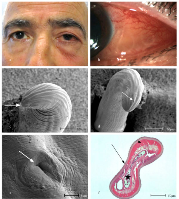
Fig. (1) (a) The 61-year-old patient with slight periorbital swelling and conjunctival injection of the left eye. (b) Close-up showing the worm coiled up in the subconjunctival space of the left eye. (c) Anterior end with mouth (arrow) in scanning electron microscopy (SEM). (d) The tail of the worm showing transverse striations and longitudinal ridges in SEM. Note spiral coiling indicating a male worm and cloaca (arrow). (e) The posterior end of the worm showing the cloaca (arrow) in SEM. (f). Transverse section of the worm with a characteristic multi-layered outer cuticle and external longitudinal ridges (arrow). Beneath the cuticle a thick muscle layer (arrowhead) is observed and internally the intestine and a single reproductive tube (star) (Haematoxylin eosin, X100 original magnification).