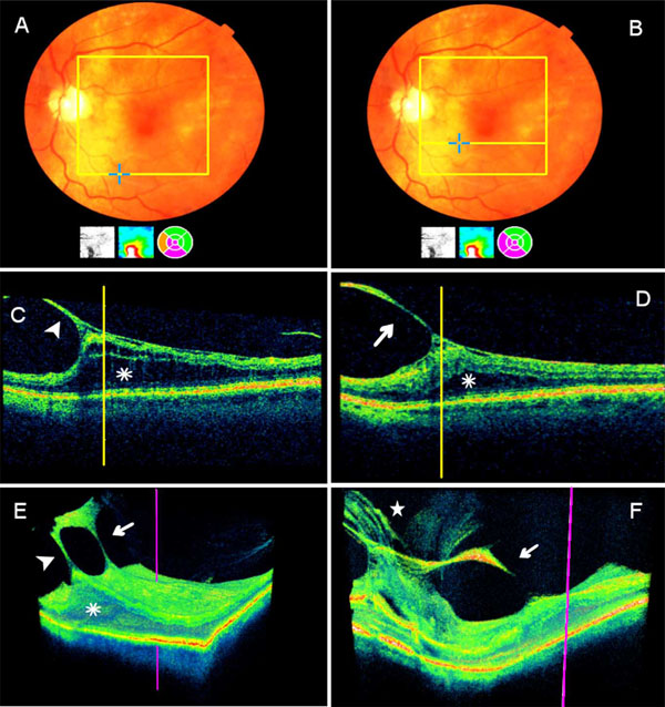
Fig. (1) Extrafoveal multifocal vitreoretinal traction and pseudophakic macular edema as detected by the spectral-domain OCT (Pt. 1). A + B. The clinical picture and macular map (the right circle map below the fundus picture) illustrate macular edema located centrally and inferonasally to the fovea (purple color). The yellow squares in A and B delineate the scanned area (6 x 6 mm). C + D. The B-mode presents two extrafoveal vitreoretinal traction membranes, marked by arrowhead in C and by an arrow in D. The manually marked vertical yellow lines indicate the traction sites; the OCT software automatically marks the anatomical locations in blue crosses, in A & B. Each of the underlying retinal edema sites (asterix) is in continuum with the central macula (seen in the map in A & B). E. 3-D image reconstruction reveals a relatively thick connection between the two vitreoretinal traction membranes associated with diffuse macular edema (asterix); the purple vertical line marks the foveal site. The arrowhead shows the inferior membrane (shown at C) and the arrow shows the superior one (shown in D). F. 3-D image reconstruction (area of 8.2 mm X 3 mm) includes the macula (the fovea is marked by the purple line), the superior traction site (arrow) and the optic nerve head (ONH; star). Multifocal vitreous membranes are associated with the ONH.