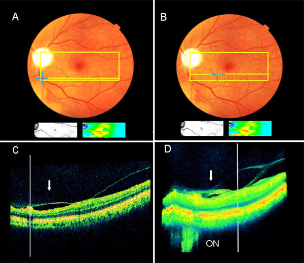
Fig. (2) Extrafoveal multifocal vitreoretinal traction associated with pseudophakic CME as detected by the SD-OCT (Pt. 2). A + B. The clinical picture and the accompanying small false-color map (below the fundus picture), as detected by 8.2mm X 3 mm scan area (the colored rectangle) illustrate macular edema (red and yellow colors). The blue crosses, one in A and one in B, demonstrate two extrafoveal vitreoretinal traction sites. One retinal edema site (in B) is shown by the false-color map to be in continuum with the central macula. C + D. The B-mode (C) and the 3-D image reconstruction (D) reveal two sites under traction by one continuous vitreous membrane (arrow). Each traction site is manually marked by a vertical white line, and its retinal location is shown automatically by yellow crosses in A & B. The optic nerve (ON) is seen in D.