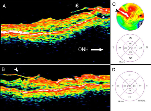
Fig. (3) Extrafoveal multifocal vitreoretinal traction associated with pseudophakic CME as detected by a time-domain OCT (Pt. 3). (A) Vitreous traction membrane (asterix) located at the papillomacular bundle area, in proximity to the ONH (not shown). An underlying localized subretinal fluid (star) and a diffuse macular edema are associated with the traction membrane. (B) A second vitreoretinal traction membrane (arrowhead) is located temporo-superiorly to the fovea; a retinal edema underlies it. (C) The 6-mm false-color and quantitative thickness macular maps disclose diffuse macular edema associated with two different tractions sites (marked by arrowhead and asterix). The central sub-field thickness is 370 µm. (D) The quantitative macular map of the normal controls (n = 12).