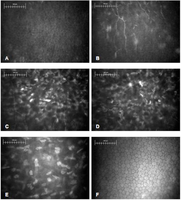
Fig. (1) Confocal images in WMS patient. Bar represents 100μm. (A) Normal basal epithelium with dark cell bodies and bright borders. (B) Sub-epithelial layer with normal nerves fibers. (C, D) Superficial stroma. Abnormally shaped, clustered keratocytes with presence of activated cells. (E) Deep stroma with normal keratocytes. (F) Corneal endothelium with normal cell morphology and density.