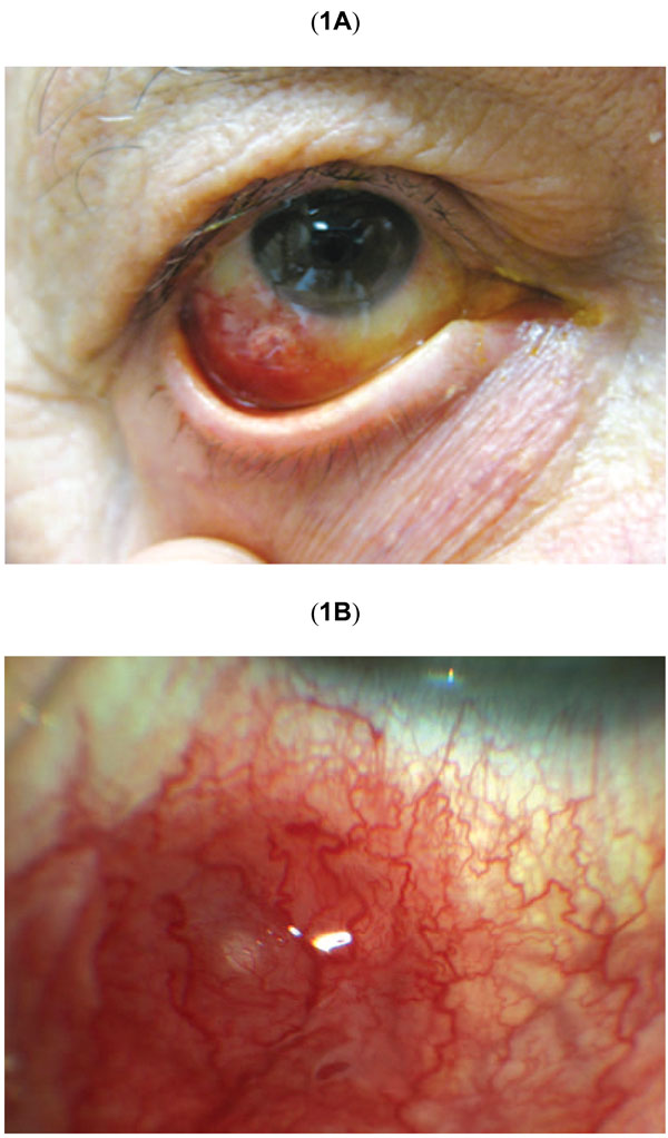
Fig. (1) (A) Color photograph shows the appearance of the right eye at presentation. There is hyperemia and edema of the inferotemporal bulbar conjunctiva. (B) Higher magnification of initial erythematous subconjunctival nodule at the site of prior intravitreal injection.