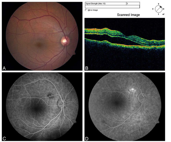
Fig. (3) (A) Color fundus photograph and (B) optical coherence tomography of the right eye showing a reduction in the amount of subretinal fluid after PDT. (C) Early and (D) late frame fluorescein angiograms showing mild dye transmission in area treated by focal laser with late staining of the laser scar in the superonasal macula.

