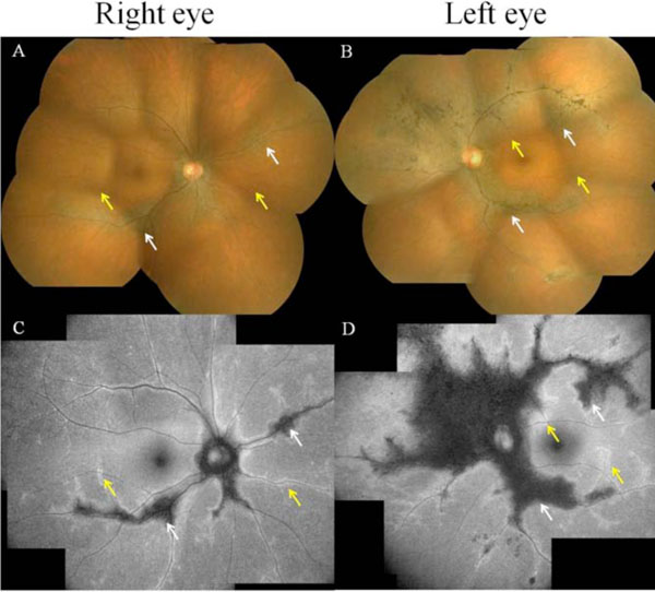
Fig. (1) A funduscopic examination reveals chorioretinal atrophy along the paravenous area in both eyes and supranasal retinal areas left eye
without macular involvement. Note also a marked bone spicule pigment clumping together with the atrophy of the left eye (A, B). Fundus
autofluorescence examination reveals geographic hypofluorescence (C, D; white arrows) along the paravenous and supranasal retinal areas.
Hyperfluorescence surrounding the hypofluorescence in the peripheral paravenous distribution (C, D; yellow arrows). These arrows are
corresponding to those shown in fundus examination.