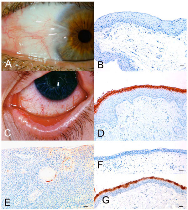
Fig. (1) A. Nasal pterygium. B. Micrograph of pterygium stained with anti-filaggrin. Bar = 100 µm. Note the complete lack of filaggrin
expression. C. Late stage of Stevens-Johnson syndrome. D. Micrograph of sample from (C). Bar = 100 µm. Note the marked binding of anti-filaggrin
to the parakeratinized superficial layers of the epithelium. E. Micrograph of conjunctiva with moderate dysplasia. Bar = 100 µm.
Only focal filaggrin expression was found. F. Micrograph of normal conjunctiva, which does not express filaggrin. Bar = 100 µm. G.
Micrograph of normal skin from same patient as in (F). Bar = 100 µm. Note the marked expression of filaggrin in the keratinized layers of
the epithelium.