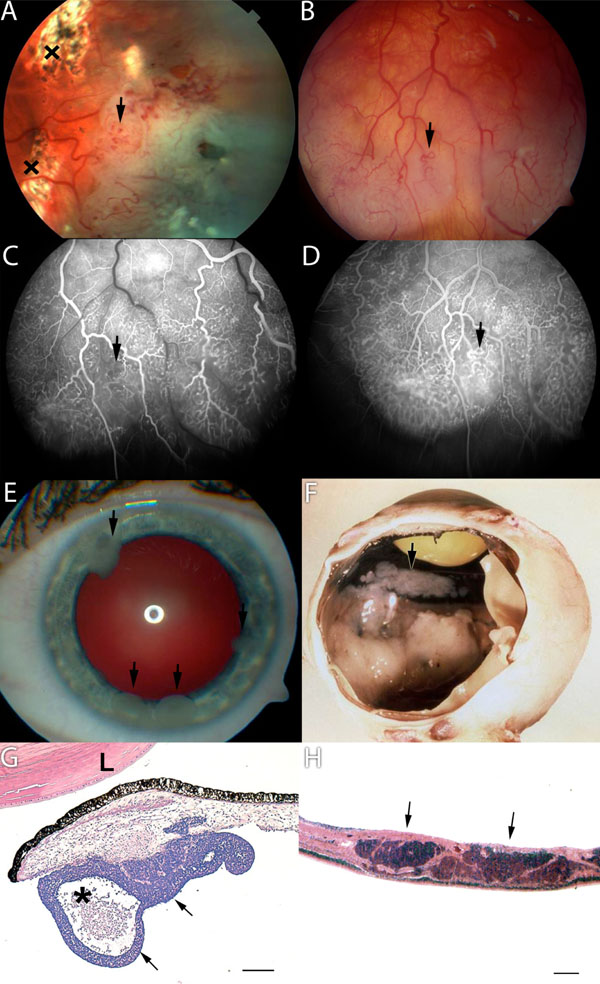
Fig. (1) (A) An elevated retinal mass covered with dilated and tortuous vessels (arrow) was observed after vitrectomy and laser coagulation
(x). (B) Five months later fundus photograph, (C) early and (D) late-phase fluorescein angiograms showed numerous abnormal intraretinal
vessels in the progressed tumor mass. Note the limited diffuse leakage in the late phase fluorescein angiogram (D). (E) Seeding of the
tumour with white lumps adherent to the front of the iris (arrows). (F) Macroscopical appearance with white tumour masses extending from
the ora serrata to the posterior pole. Multifocal greyish tumour seeding was covering the back of the iris and the pars planitis corpus ciliare
(arrow). (G) Micrograph of tumour satellite on anterior iris surface (arrows). The lesion has a cystic appearance because of the central
necrotic space (asterisk). L = lens, hematoxylin and eosin stain, bar 500 µm. (H) Micrograph of retina with intraretinal tumour infiltration.
Note the nearly complete lack of endo- and exophytic tumour growth. Hematoxylin and eosin stain, bar 100 µm.